Figure 4.
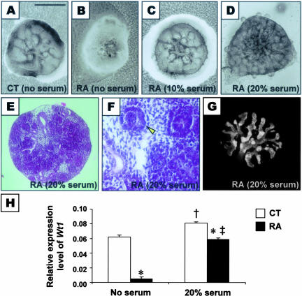
Metanephric explants cultured in vitro for 5 days. A–D: Morphology of explanted metanephroi from control (CT) or RA-treated (RA) embryos cultured in serum-free medium (A and B), or medium supplemented with 10% (C) or 20% serum (D). E and F: Histological sections of RA-treated metanephric explants cultured in 20% serum showed that the rudiments had undergone differentiation, with formation of condensates, primitive tubules, and avascular glomeruli (arrowhead). G: Calbindin immunostaining of UB branch epithelium showed multiple branching in metanephric explants from RA-treated embryos after culture in 20% serum. H: Real-time quantitative RT-PCR analyses of expression levels of Wt1 relative to β-actin in metanephroi from control and RA-exposed embryos at 2 days after culture in serum-free medium or medium supplemented with 20% serum [*, P < 0.0001, CT versus RA in the same culture condition; †, P < 0.005, CT (no serum) versus CT (20% serum); ‡, P < 0.0001, RA (no serum) versus RA (20% serum); Student’s t-test]. Scale bar: 0.5 mm (A–D, G), 0.35 mm (E), 1.4 mm (F).

