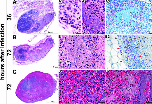
Figure 6.
Progression of histopathology in the primary bubo. Sections of proximal lymph nodes collected at 36 (A) and 72 (B and C) hours after intradermal inoculation of ∼300 Y. pestis were stained by H&E (A, A1 and A2; B, B1; and C, C1 and C2) or by Naphthol AS-D chloroacetate esterase, which stained PMNs (A3 and B2). Gray arrowheads indicate individual extracellular bacteria (A1) or bacterial aggregates, which appear as fields of blue (A3) or purple (A, A2, B1, C1, and C2). PMNs stain red by chloroacetate esterase (A3 and B2) and were localized at the periphery of the lymph node (delimited by red arrowheads in B2) and surrounded the bacterial aggregates. PMNs in H&E sections are indicated by black arrowheads (A1 and A2). Fibrin appears as deposits of pink color, indicated by green arrowheads (B, B1, C, C1, and C2). Blue arrowheads show cellular debris and condensed, degraded nuclei, indicative of necrosis or apoptosis (B1 and C2). Unlabeled scale bars, 20 μm.

