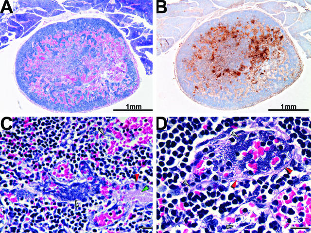
Figure 7.
Histopathology of distal lymph node infected by hematogenous spread (secondary bubo). Section of an infected right maxillary lymph node was stained by H&E (A, C, and D) or by IHC using Y. pestis-specific antibody (B). Bacteria (stained brown by IHC) colocalize with areas of hemorrhage (stained red by H&E), which appeared to be the consequence of vasculitis. Infected, disrupted blood vessels, indicated by red arrowheads (C and D) contain intra- and perivascular bacterial aggregates (dark purple areas indicated by gray arrowheads), fibrin deposits (pink areas indicated by green arrowheads), and hemorrhage. Unlabeled scale bars, 20 μm.

