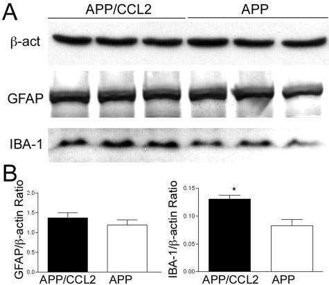
Figure 8.
GFAP and IBA-1 expression in young mouse brains. A: Protein extracts (30 μg/lane) from the frontal cortex of APP/CCL2 and APP mice at 5 months of age (n = 3 per group) were subjected to immunoblotting for β-actin, GFAP, and IBA-1. B: Quantification of immunoreactive bands in A. *, P < 0.05 versus APP as determined by Student’s t-test.

