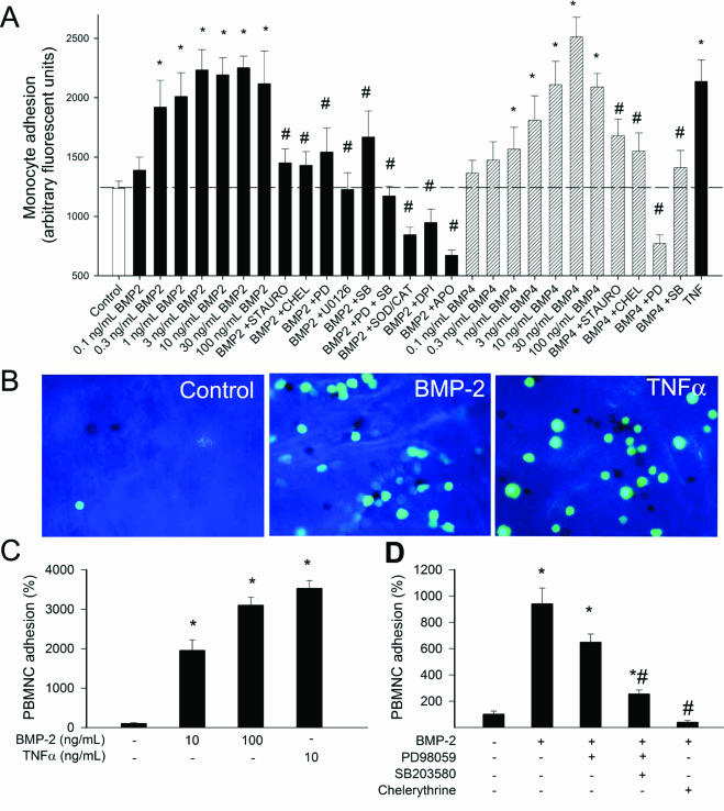
Figure 6.
A: Results of monocyte adhesion assay (see Materials and Methods). Treatment of primary human CAECs with increasing concentrations of BMP-2 and BMP-4 (2 hours) significantly increased the adhesion of fluorescently labeled PMA-stimulated monocytes. TNF-α (10 ng/ml) was used as positive control. The effects of 10 ng/ml BMP-2 and BMP-4 were also assessed after pretreatment with inhibitors on p42/44 MAP kinase (10 μmol/L PD 98059, 10 μmol/L U0126), p38 MAP kinase [10 μmol/L SB203580, PKC (10 μmol/L chelerythrine (CHEL), 10 μmol/L staurosporine (STAURO)], NAD(P)H oxidase (10 μmol/L DPI, 3 × 10⁻⁴ mol/L APO), and the free radical scavenger SOD plus catalase (200 U/ml). Data are mean ± SEM (n = 8 for each group). *P < 0.05 versus untreated control. #P < 0.05 versus BMP treatment. B: Representative fluorescent images showing the effect of BMP-2 and TNF-α treatments (2 hours) on the adhesion of activated monocytes (green fluorescence) to the endothelium of rat aortic segments (en face preparation). C: Summary bar graphs are shown (data are normalized to control mean values). D: Effects of PD 98059, SB203580, and chelerythrine on BMP-2 induced monocyte adhesion to the aortic endothelium. Data are mean ± SEM. *P < 0.05 versus control, #P < 0.05 versus BMP-2 treatment. Original magnifications, ×40.

