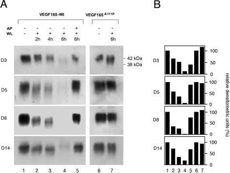
Figure 4.
Stability of VEGF165A111P protein is increased in db/db wound tissue lysate. A: VEGF165-Wt (lanes 2 to 5) or VEGF165A111P (lanes 6 and 7) protein was incubated with wound tissue lysates (WL) harvested from db/db mice at indicated time points after wounding (days 3 to 14), for increasing time periods as indicated (2 to 6 hours). α2-Antiplasmin (AP) could partially inhibit VEGF165-Wt degradation (lane 5). Degradation of VEGF165 variants was monitored by SDS-PAGE under nonreducing conditions. B: Degradation of VEGF165 variants incubated in wound tissue lysates was monitored by analyzing the intensity of the 42-kd signal by scanning densitometry and is presented as a percentage of the signal intensity before incubation into wound tissue lysate (A, lane 1). Lane numbers in B correspond to the lane numbers presented in A.

