Figure 6.
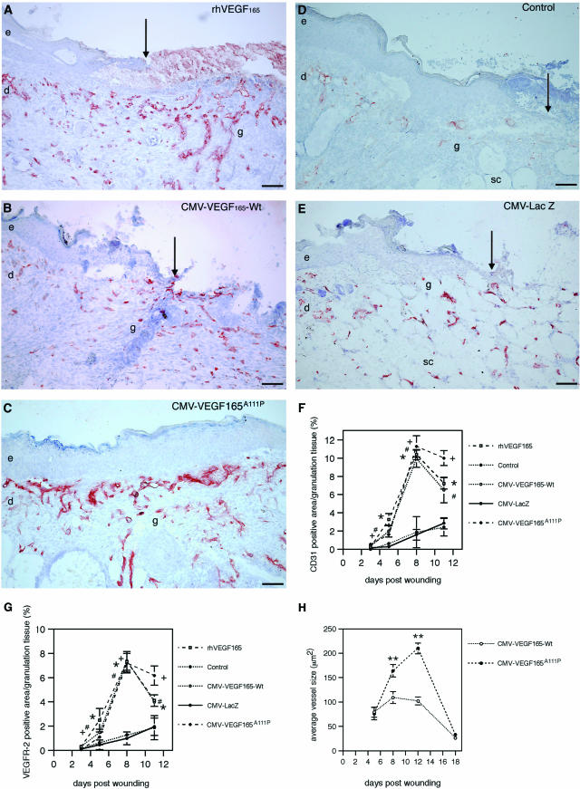
Vascular density is increased in VEGF165-treated wounds in db/db mice. Representative immunofluorescent staining demonstrates CD31-stained blood vessels (red) 8 days after wounding. Pronounced increase of microvascular density in granulation tissue of wounds treated topically with rhVEGF165 protein (A) or transfected with CMV-VEGF165-Wt (B) or CMV-VEGF165A111P (C) expression constructs. Note that mutant-transfected wound shows a hyperproliferative epithelium, which just reached complete re-epithelialization. Significantly reduced granulation tissue and microvascular density in granulation tissue of methylcellulose-treated (control) (D) or CMV-LacZ-transfected (E) wounds. Note that due to the lack of granulation tissue in control and CMV-LacZ-treated wounds the subcutaneous fat tissue, which is abundant in db/db mice, is visible beneath the epithelial wound edges. Blood vessels stained in CMV-LacZ-treated wounds are localized in the subcutaneous fat tissue. Time course of computer-assisted morphometric analysis of CD31-positive cells (F) or VEGFR-2-positive cells (G) within the granulation tissue. H: Average vessel size is increased in CMV-VEGF165A111P- versus CMV-VEGF165-Wt-transfected wounds. Wounds are n = 8/time point for each treatment modality; data are expressed as mean ± SD (rhVEGF165 versus control, *P < 0.05; CMV-VEGF165-Wt versus CMV-LacZ, #P < 0.03; CMV-VEGF165A111P versus CMV-LacZ, +P < 0.003; CMV-VEGF165-Wt versus CMV-VEGF165A111P, **P < 0.03). e, epidermis; d, dermis; sc, subcutaneous tissue; g, granulation tissue. Arrows indicate epithelial wound edge. Scale bars, 100 μm.

