Figure 7.
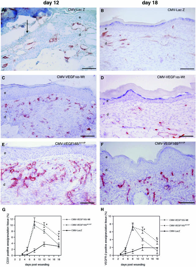
Persistence of increased microvascular density in CMV-VEGF165A111P-transfected wounds. Representative immunofluorescent staining demonstrates CD31-stained blood vessels (red) 12 and 18 days after wounding. A and B: Poorly vascularized granulation tissue in CMV-LacZ-transfected wounds. Note that blood vessels stained in CMV-LacZ-treated wounds are predominantly localized in the subcutaneous fat tissue. Pronounced induction of neovascularization in granulation tissue of wounds transfected with CMV-VEGF165-Wt (C) or CMV-VEGF165A111P (E) expression constructs 12 days after wounding. D: Significantly reduced microvascular density in granulation tissue of CMV-VEGF165-Wt-transfected wounds at day 18 after wounding. F: Increased microvascular density in granulation tissue of CMV-VEGF165A111P-transfected wounds persists until day 18 after wounding. G: Computer-assisted morphometric analysis of CD31-stained (G) or VEGFR-2-stained (H) wound sections revealed a significantly prolonged persistence of capillaries in granulation tissue of CMV-VEGF165A111P-transfected wounds at days 12 and 18 after wounding when compared with CMV-VEGF165-Wt-transfected wounds. Wounds are n = 8/time point for each treatment modality; data are expressed as mean ± SD (CMV-VEGF165-Wt versus CMV-VEGF165A111P, *P < 0.003). e, epidermis; d, dermis. Scale bars, 100 μm.

