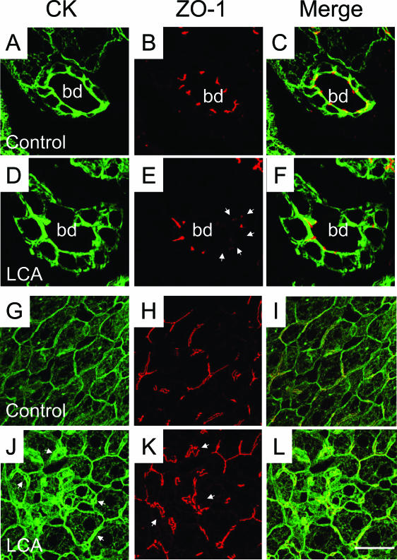
Figure 6.
Tight junctions are altered in LCA-fed mouse liver. Double-immunofluorescence labeling for CK8/18 (in green) and the tight junction protein ZO-1 (in red) in control liver (A–C and G–I) and the liver from a 4-day LCA-fed mouse (D–F and J–L). B and C: ZO-1 staining of bile ducts in control diet-fed mice shows a distinct pattern outlining the cell contacts between BECs. E and F: In contrast, the liver of a 4-day LCA-fed mouse shows strikingly altered tight junctions morphologically characterized by partially disrupted and missing ZO-1 staining (arrowheads). Note also that BECs still form an intact epithelial monolayer (D and F), suggesting that the observed missing ZO-1 staining is not related to BEC necrosis. G–I: Staining of the intermediate filament network and tight junctions of hepatocytes in control diet-fed mice. J: The liver of a 4-day LCA-fed mouse shows a significantly increased density of the cytokeratin intermediate filament (CK-IF) network with a pronounced pericanalicular sheath (arrowheads). K: Strikingly altered tight junctions between hepatocytes in 4-day LCA-fed mouse liver morphologically characterized by elongation and distortions of the ZO-1 staining pattern (arrowheads). bd, bile duct. Bar = 10 μm.

