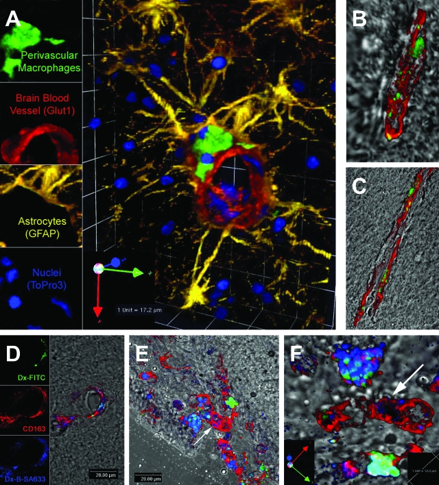
Figure 3.
Localization and turnover of CD163+ perivascular and meningeal macrophages labeled by dextran amine dye injected into the CSF of live monkeys. Confocal microscopy studies of brain sections from normal noninfected rhesus macaques injected intracisternally with fluorescent dextran amine dye (Fluoro-Emerald; green), showing localization of dye-labeled CD14+CD163+ perivascular macrophages in the perivascular space (A, B, and C). A: Dextran amine dye specifically labels CNS perivascular macrophages in normal noninfected rhesus macaques. Perivascular macrophages (green) adjacent to a CNS microvessel (Glut-1; red). Astrocytes (GFAP; yellow) and cell nuclei (ToPro3; blue). B: Dye-labeled perivascular macrophages (green) are CD163 positive (red) in CNS of a normal noninfected rhesus macaque. C: Dye-labeled perivascular macrophages (green) are CD14 positive (red). Confocal images from a noninfected animal subjected to consecutive injections of Fluoro-Emerald (green) followed by biotinylated dextran amine (BDA-10,000; visualized with streptavidin-Alexa Fluor 633; blue) 7 days later (D, E, and F). The animal was sacrificed at 24 hours after BDA-10,000 injection. D: Sequential labeling of perivascular macrophages by consecutive injections of Fluoro-Emerald (green; top leftpanel) followed 7 days later by biotinylated dextran (blue; bottom leftpanel) detects double-positive (green and blue) and single-positive (blue) perivascular macrophages, both of which are CD163 positive (red). The sequential labeling identifies two populations of CD163+ perivascular macrophages in perivascular spaces: perivascular macrophages that do not turnover in a 7-day period (green-blue) and perivascular macrophages that have recently arrived in the CNS (blue only). E: Sequential labeling by consecutive injections of Fluoro-Emerald (green) followed 7 days later by biotinylated dextran (blue) demonstrates double-positive (green-blue) and single-positive (blue) meningeal macrophages, all of which express CD163. The majority of cells are double positive, with few single-positive cells that have recently arrived. F: Magnification of E. High-power magnification view demonstrates double-positive (green-blue) and single-positive (blue) meningeal macrophages in the same field of meninges (horizontally flipped rear view image of E). Arrows point to the identical CD163+ meningeal macrophage that does not contain Fluoro-Emerald but BDA-10,000 (blue only).

