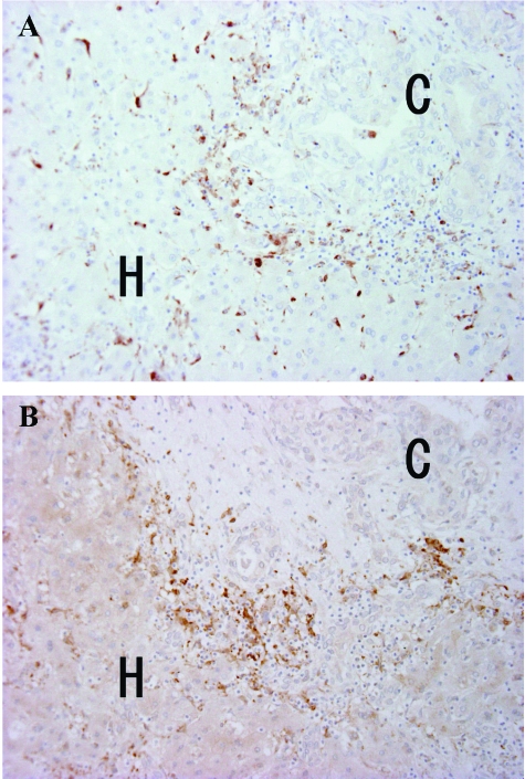
Figure 1.
A and B are serial sections and correspond to almost the same area of ICC. A: There are many CD68-positive macrophages infiltrating at the interface of the ICC (C) and surrounding liver (H). Kupffer cells in the surrounding liver are also strongly positive. Immunostaining for CD68 counterstained by hematoxylin. B: There are many TNF-α-positive mononuclear cells infiltrating at the interface of the ICC (C) and surrounding liver (H). A majority of them correspond to infiltrating macrophages. Kupffer cells in the surrounding liver are also strongly positive. Immunostaining for TNF-α counterstained by hematoxylin. Original magnifications, ×200.

