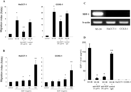
Figure 7.
A: Migration of cultured ICC cells increased on co-culture with WI-38 fibroblasts in the lower chamber for 48 hours (migration of HuCCT-1 cells increased 5.5-fold and that of CCKS-1 increased 20.8-fold, compared with the control). When anti-SDF-1 neutralization antibody was added to the culture medium of ICC cells co-cultured with WI-38 fibroblasts, the increase in migration of ICC cells was down-regulated (from 5.5- to 1.7-fold in HuCCT-1 and from 20.8- to 34-fold in CCKS-1, respectively). Migration of ICC cells (HuCTT-1 or CCKS-1) co-cultured with WI-38 cells on control IgG was 4.85 ± 0.93 and 19.42 ± 2.14, almost similar to that of ICC co-cultured with WI-38 cells. *P < 0.05 versus control. **P < 0.05 versus HuCCT-1 + WI-38 fibroblasts or CCKS-1 + WI-38 fibroblasts. Migration index is the ratio of the number of migrated cells in an experimental group/the number of migrated cells in control group. The data are provided as the mean ± SD. The experiments were performed five times. B: Migration of cultured ICC cells of HuCCT-1 increased when 1, 10, and 100 ng/ml SDF-1 was added in the lower chamber [migration index is 1.3-, 1.4-, and 5.3-fold, respectively, compared with the control (ICC cells alone are cultured)]. Migration of CCKS-1 increased when 10 and 100 ng/ml of SDF-1 was added (migration index is 1.7- and 4-fold, respectively). *P < 0.05 and **P < 0.01 versus control. The number of migrated cells was counted in 10 medium power fields (×20). The data are provided as the mean ± SD. Migration assay was performed five times in each experiment. C: RT-PCR shows that SDF-1 mRNA is detected in cultured WI-38 fibroblasts. SDF-1 mRNA is not detected in cultured HuCCT-1 or CCKS-1 cells. D: ELISA shows that SDF-1 protein is present in the supernatant of cultured WI-38 fibroblasts (172.3 ± 22.2 pg/ml), but it was not detected in the supernatant of cultured HuCCT-1 or CCKS-1 cells. The addition of 5 ng/ml and 50 ng/ml of anti-SDF antibody in the supernatant of the cultured WI-38 fibroblasts almost eliminated SDF-1 (8.8 ± 4.0 pg/ml and 16.3 ± 3.1 pg/ml), whereas SDF-1 level in the supernatant of WI-38 cells treated with control IgG was 141.0 ± 7.0 pg/ml, similar to that of WI-38 cells alone. *P < 0.01 versus WI-38 cells alone and WI-38 cells + control IgG. **P > 0.05 versus WI-38 cells. The data are provided as the mean ± SD, and the SDF-1 level was measured three times in each experiment.

