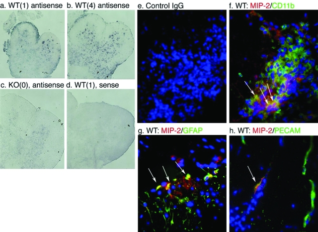
Figure 4.
CXCL2 is produced by astrocytes and vascular endothelium of the CNS as well as CD11b+ cells from the inflammatory infiltrate early in the disease course. In situ hybridization analysis of spinal cord sections of irradiated WT (a, b, and d) and TNFR1-null (c) mice 6 days after transfer of 5 × 10⁶ Thy 1.1 cells using digoxigenin-labeled anti-sense (a–c) and sense (d) riboprobes. Clinical scores of animals are shown in parentheses. Immunohistochemical analyses of CXCL2 (red) expression within CD11b- (green) (f), GFAP- (green) (g), and PECAM-expressing cells (green) (h). Nuclei have been counterstained with DAPI. Arrowheads: Co-localization (yellow) of CXCL2- and CD11b- (f), GFAP- (g), and PECAM- (h) positive cells. Arrows: MIP-2-negative, PECAM-positive endothelium. e: Control IgG does not demonstrate any specific staining. Analyses performed on sections from six animals, n = three mice per group. Original magnifications, ×350.

