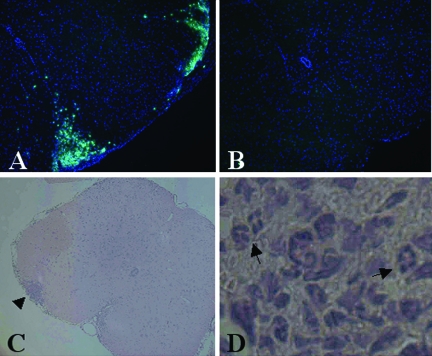
Figure 5.
Recruitment of peripheral GR1+ cells to the CNS is blocked in TNFR1-null mice. Th1-skewed Thy1.1 WT CD4+ MOG-specific T cells (5 × 10⁶) were injected intravenously into sublethally irradiated (450 R) 6- to 8-week-old Thy1.2 B6 WT and TNFR1-null recipients. Frozen sections of lumbar-sacral spinal cord from day 6 WT, CS = 1.5 (A), and day 6 TNFR1-null, CS = 0 (B) were immunostained for GR-1 (green) and counterstained with DAPI (blue). C and D: H&E section of lumbar-sacral spinal cord from day 7 WT, CS = 1, shows a focus of inflammatory infiltrate (arrowhead) invading the parenchyma, which contains many neutrophils (arrows). Original magnifications: ×65 (A–C); and ×260 (D).

