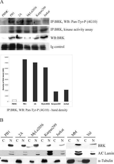
Figure 2.
Activation status and subcellular localization of Brk in selected T- and B-cell populations. A: Kinase activity assay to determine activation status of Brk as detected by its autophosphorylation and Western blot to examine protein phosphorylation using anti-phosphotyrosine antibody (4G10). The protein phosphorylation is shown also schematically by depicting densitometric analysis data. B: Western blot: Brk protein expression in fractionated nuclei and cytoplasm with A/C lamin (nuclear protein) and a-tubulin (cytoplasmic protein) serving as controls.

