Figure 3.
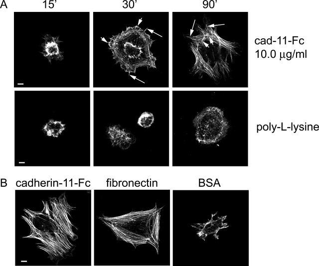
Cadherin-11 engagement stimulates actin polymerization and actin cytoskeletal reorganization. A: FLSs were plated on protein-coated coverslips as indicated in the absence of serum for the indicated times, fixed, permeabilized, stained for F-actin (phalloidin-Alexa 488, green), and analyzed by confocal microscopy. Note time-dependent cell spreading. After 30 minutes the cells plated on cadherin-11-Fc fusion protein demonstrated radial spikes (middle, long arrows) and lamellipodial protrusions (middle, short arrows). After 90 minutes pronounced cell spreading was accompanied by the formation of radial actin fibers (right, long arrows) and circumferential actin fibers (right, short arrows). In contrast, in FLSs plated on poly-l-lysine, radial actin fibers and lamellipodial protrusions were not detectable. B: FLSs were plated on cadherin-11-Fc fusion protein-, fibronectin-, or BSA-coated coverglasses. Cadherin-11-Fc induced pronounced cell spreading and actin fiber formation in a similar way as fibronectin, whereas BSA failed to induce cell spreading. Scale bars, 10 μm.

