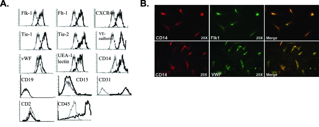
Figure 2.
Flow cytometric analyses of CE-EPC phenotype demonstrate that the majority expressed markers of both endothelial and monocyte lineages. A: Representative flow cytometry histograms of CE-EPCs after 7 days of culture obtained from PB-MNCs. Parameters for specific antibody staining (thick line) set according to the isotype controls. B: Co-expression of CD14 and endothelial markers (Flk1 or vWF) was detected by immunofluorescent analysis on day 7 CE-EPCs derived from PB (top) or BM (bottom). Magnifications, ×200.

