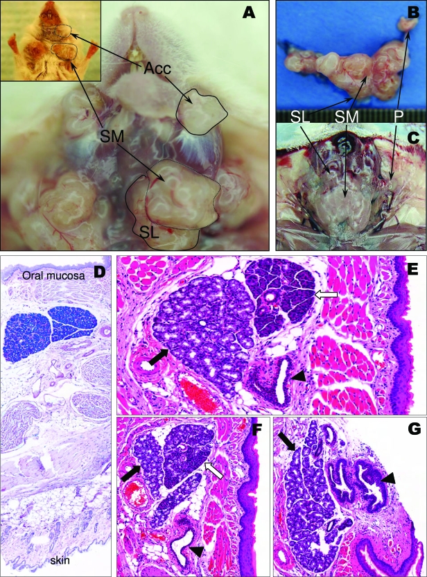
Figure 1.
A: Gross anatomy of the salivary glands in a K5-tet-on/tet-o-ras animal. All glands are grossly distorted and enlarged showing a multinodular surface (SM: submaxillary, SL: sublingual). The accessory buccal glands (Acc) are also enlarged. The inset shows the glands already visible as tumor-like structures protruding through the neck skin. The abnormal gross anatomy is better depicted in the dissected gland (B) surface (SM: submaxillary, SL: sublingual, P: parotid); C: Relatively flat end even surface of normal salivary glands for comparison. D–G: Normal histology of the accessory salivary glands. These are microscopic mixed, mucous, and serous glands (inset, ductal structure), inconspicuous unless enlarged, located beneath the superficial muscular layer close to the buccal mucosa. D: Location of the glands in relation to the oral mucosa and the skin (Alcian Blue, 64×). E: Serous (white arrow) and mucinous (black arrow) lobules are separated at this level by a thin connective wall. In deeper sections both components coalesce (F), and the serous component disappears in even deeper sections (G). Excretory ducts are indicated by an arrowhead. (Different amplifications of pictures taken at 10× are shown.)

