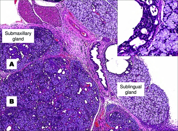
Figure 2.
Dysplastic changes with solid squamous metaplastic proliferation (A) and adenosis (B) are evident in the submaxillary gland. The parenchyma of the sublingual gland seems unaffected, but the extralobular ducts show focal intraductal cribiform proliferation (inset). Magnifications: ×40 and ×100 (inset).

