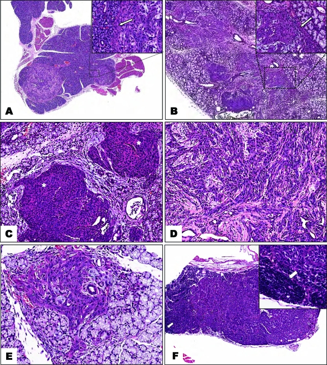
Figure 4.
A: This multifocal squamous cell carcinoma arises in an otherwise unaffected submaxillary gland; no signs of hyperplasia or dysplasia are observed. The inset shows a distinct limit between neoplasia and normal tissue (arrow). B: Multifocal spindle-cell carcinomas in a sublingual gland arising from ductal structures; the inset depicts the border between the carcinoma and the normal tissue; a fibrous pseudocapsule with inflammatory mononuclear cells is seen (arrow). C: Moderately differentiated multifocal squamous cell carcinoma arising in a submaxillary gland; the tumors form solid masses that infiltrate the surrounding stroma. Distorted mucous acini are seen in the periphery. D: Infiltrating spindle-shaped carcinoma, with a desmoplastic stroma. E: Early carcinoma arising in a dysplastic sublingual gland. Note that, although the lesion is small, all of the cells display malignant features; a rapid transition to invasion characterized these tumors, with no evident dysplasias. F: Metastatic squamous cell carcinoma in a cervical lymph node; the arrows indicate remnants of the lymph node parenchyma. Magnifications: ×20 (A); ×30 (B); ×45 (C–F); and ×100 (insets).

