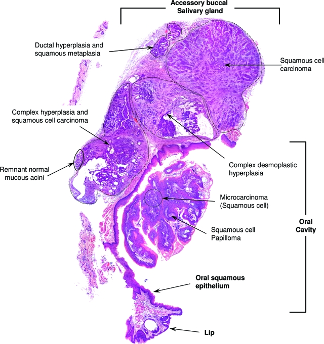
Figure 5.
Panoramic view of a buccal accessory salivary gland with a wide spectrum of histological changes from hyperplasia to carcinoma, showing its relations with the oral cavity. Note that, unlike the extraorbital lacrimal glands, these structures contain mucous acini, remnants of which are shown. A squamous papilloma of the oral epithelium that has an area of invasive carcinoma can also be seen. Magnification: ×40.

