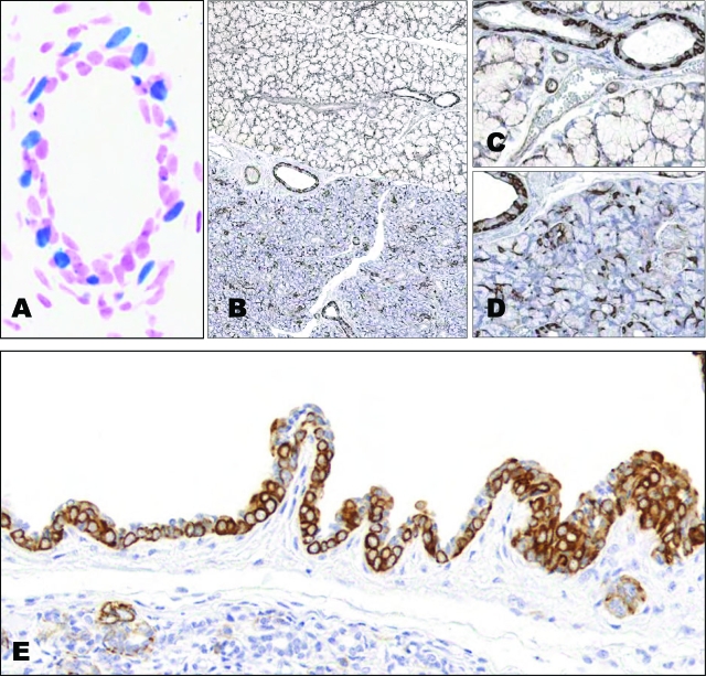
Figure 6.
A: β-Galactosidase staining; isolated ductal cells are positive. B: Immunostaining for keratin 5 in sublingual (upper figure) and submaxillary (lower figure) glands reveals positive staining in most ductal cells as well as in myoepithelial cells. C: Higher magnification of K5 expression in the sublingual gland. D: Submaxillary gland immunoreacted with K5. E: K5-tet-on/tet-o-ras mice. K5 is expressed in all proliferating cells in a hyperplastic excretory duct. Magnifications: ×40 (B); ×60 (C–E); and ×120 (A).

