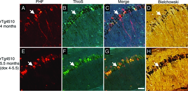
Figure 5.
Persistent neurofibrillary pathology after transgene suppression. A and E: PHF1-positive accumulations in neurons persist after 6 weeks of transgene suppression. B and F: These lesions are also Thioflavine S (ThioS)-positive as can be seen in the merged images (C and G). D and H: Bielchowski silver staining shows that these same lesions are argyrophilic, indicating abnormal confirmation as well as hyperphosphorylation persists after transgene suppression. Arrows indicate cells positive for PHF1, Thioflavine S, and Bielchowski stains. Scale bars, 100 μm.

