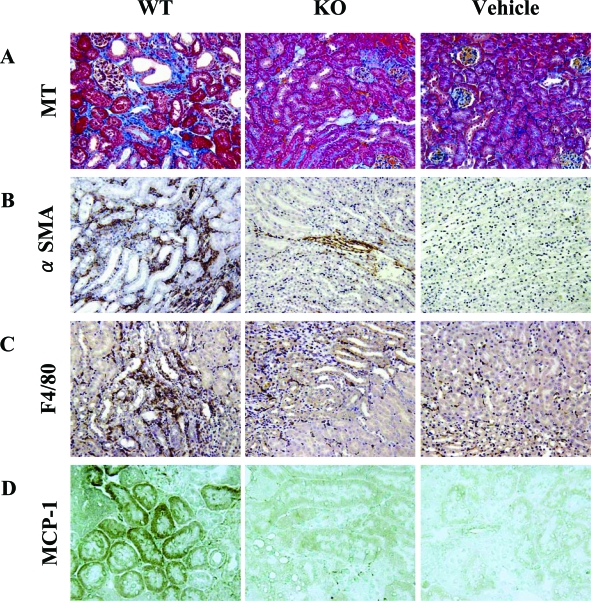
Figure 6.
Histological findings at day 14 stained with Masson’s trichrome (A), immunohistochemistry using antibody to α-SMA (B), antibody to F4/80-positive macrophage (C), and immunohistochemical analyses using antibody to MCP-1 with frozen sections. Vehicle-treated PAFR-KO and PAFR-WT showed identical results. Therefore, only PAFR-WT mice results are shown. Original magnifications: ×100 (A); ×200 (B, C); ×400 (D).

