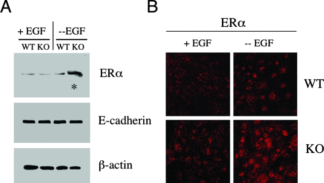
Figure 6.
Cav-1-deficient mammary epithelial cells up-regulate the ERα under conditions of growth factor deprivation. A: Western blot analysis. WT and Cav-1-deficient mammary epithelial cells were isolated and cultured in Matrigel in the presence or absence of EGF. Under these conditions, mammary epithelial cells are able to form 3-D structures that closely resemble mammary acini. Lysates were then prepared from day 16 acini and subjected to Western blot analysis with anti-ERα specific antibodies (MC-20; Santa Cruz Biotechnology). Note that, under conditions of growth factor deprivation (in the absence of EGF), Cav-1 KO acini display a substantial increase in ERα expression levels (see asterisk). Equal loading was assessed by immunoblotting with an E-cadherin antibody. B: Immunofluorescence. To evaluate ERα localization and expression, WT and Cav-1 KO mammary epithelial cells were grown on glass coverslips for 6 days and cultured in either the absence or presence of EGF overnight. Then, cells were fixed and subjected to immunofluorescence analysis with an antibody directed against ERα. Note that, in the absence of EGF, Cav-1 KO mammary epithelial cells exhibit increased ERα expression levels, as compared to their WT counterparts. We also observed intense ERα nuclear staining in Cav-1-deficient mammary epithelial cells, consistent with ERα receptor activation.

