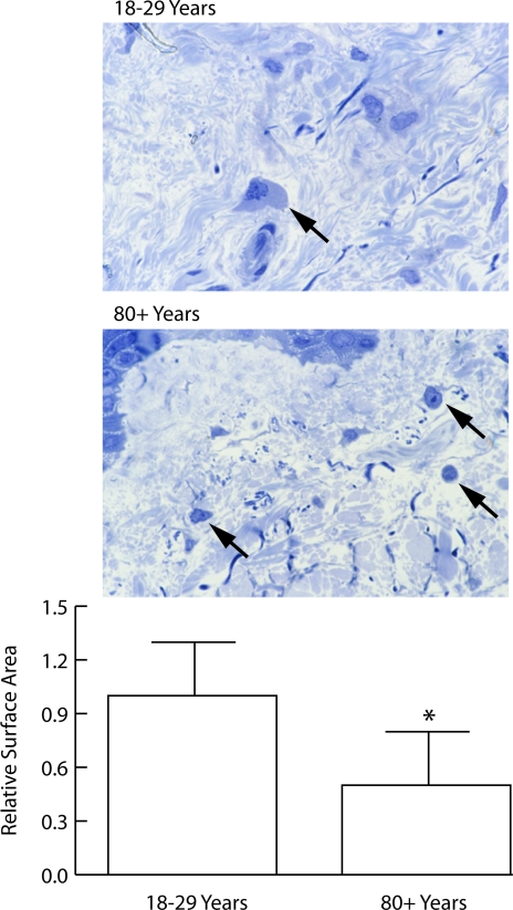
Figure 4.
Shape of fibroblasts in the papillary dermis of sun-protected hip skin from young and old individuals (1-μm toluidine blue-stained sections from glutaraldehyde-fixed, plastic-embedded tissue). Top panel: Cells in young skin are flattened, and cytoplasm and nucleus are visible (arrow). Cells are embedded in matrix. Cells in old skin appear round, and only the nucleus and a small amount of cytoplasm are visible (arrows). Bottom panel: Surface area measurements were made quantitatively as described in Materials and Methods. Values represent mean cross-sectional surface area ± SEM, based on 160 cells in sun-protected skin from six young individuals and 57 cells in sun-protected skin from six old individuals. Statistical significance was determined using Student’s t-test (two-tailed). *P < 0.01 (magnification, ×240).

