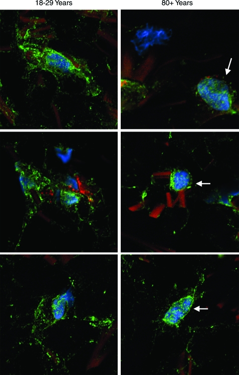
Figure 6.
Adhesion-site protein expression in sections of healthy sun-protected hip skin from young and old individuals. Tissue sections (OCT-embedded frozen) were stained using antibody to vinculin and concomitantly with phalloidin (actin stain) and DAPI (nuclear stain) as described in Materials and Methods. After staining, cells were examined by confocal fluorescence microscopy. Cells are identified by their blue (DAPI)-stained nuclei. Bright green punctate fluorescence identifies vinculin. In the 18- to 29-year-old skin samples, vinculin can be seen at a distance from the nucleus, and in many areas, the vinculin appears to be in close apposition to collagen fibers. In the 80+-year-old skin, blue-stained nuclei are apparent, but there is less vinculin than in the young skin samples. Where intense focal staining is evident, it is surrounding the nucleus (arrows). Away from the nucleus, staining is more diffuse than seen in cells from young skin. The sections presented are representative of young and old sun-protected skin from six individuals, respectively. In both young and old skin, collagen fibers are apparent by their dull orange fluorescence (magnification, ×1200).

