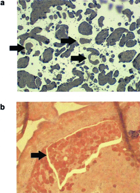
Figure 1.
Laser capture microdissection of trophoblast cells. a: Photomicrograph of placental villi from the placental basal plate region (magnification: 20×). b: Photomicrograph of extravillous trophoblast column from the placental basal plate region (magnification: 100×). Extravillous trophoblast cells were immunostained with anti-cytokeratin-18 antibodies for identification. Arrows indicate regions where cells were captured.

