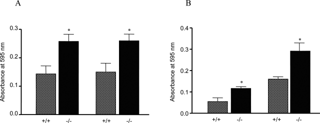
Figure 11-6870.
Lymphocyte proliferation. Peripheral lymphocytes were collected from lymph nodes (mandibular and axillary regions) from 6-month-old mice. CD4+ cells were isolated using BD IMagnet. Lymph node cells (A) or CD4+ lymphocytes (B) were seeded at a density of either 4 × 10⁴ (left) or 1 × 10⁵ (right). The cells were stimulated with anti-CD3 antibodies for 48 hours. Proliferation was measured with the MTT kit. ░⃞, Nrf2+/+; ▪, Nrf2−/−. Data represent means ± SD from three samples. *P < 0.05.

