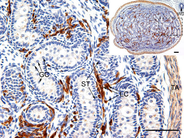
Figure 8.
Immunolocalisation of WNT4 in the testis of the tammar at day 45pp. WNT4 immunostaining was present in the Leydig cells and the tunica at day 45pp, but it was absent from germ cells and Sertoli cells and Wolffian duct (WD). The dashed square on the low-power image (inset) is the area shown in high-power. Germ cells (GC), Sertoli cells (ST), Leydig cells (LC) and tunica albuginea (TA). Scale bars = 100 μmm.

