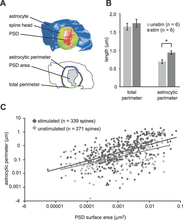
Figure 4. Whisker Stimulation Increases the Astrocytic Participation at the Bouton–Spine Interface.
(A) Shows a 3D reconstruction of a spine head (green), its PSD (red), and the associated astrocyte (blue). The orientation of the structure shows the region occupied by the axonal bouton (removed). The line drawing below shows the parameters measured: the total perimeter of the interface between the bouton and the spine, and the part of this perimeter that is occupied by the astrocyte, the astrocytic perimeter.
(B) Stimulation did not change the degree of contact between bouton and spine, measured by the total perimeter (p > 0.5). However, the amount of the perimeter occupied by the astrocyte was significantly increased (p < 0.0001), using mean values per animal.
(C) Correlation between the length of the perimeter that is occupied by astrocytic membrane and the PSD surface area on spines in unstimulated (light grey diamonds, n = 271; p < 0.001, R2 = 0.68) and stimulated neuropil (dark grey diamonds, n = 340; p < 0.001, R2 = 0.73).

