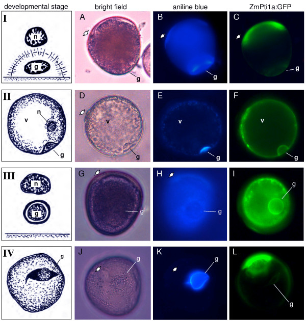
Figure 7.
ZmPti1a:GFP localization during pollen mitosis I. Bright field, decolorized aniline blue staining and GFP epifluorescence of representative ZmPti1a:GFP transgenic pollen grains at four stages of pollen development. (I) Callose stage; (II) Compact callose stage; (III) Circular shaped prophase generative nucleus; (IV) Spindle shaped prophase generative nucleus. At this stage, the generative cell is still encased by callose and becomes spindle shaped. Later on, callose disappears and the generative cell immediately undergoes a second mitotic division resulting in trinucleate mature pollen. Stages I and III redrawn from [26]; stage II drawn according to [61]; stage IV redrawn from [62]; g = generative cell; n = vegetative nucleus; v = vacuole; A/B, G/H and J/K, show the same pollen grains, respectively.

