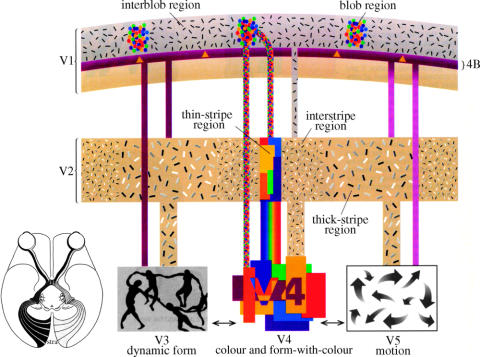
Figure 13.
A diagrammatic representation of areas V1 and V2 and their compartments, as well as three areas of the visual brain. Layers 2 and 3 of V1 are characterized by metabolically active ‘blobs’ in which wavelength-selective cells are concentrated and, between them, the interblobs, which contain the orientation-selective cells. Directionally selective cells are concentrated in layer 4B. These compartments project in an orderly way to specific compartments of V2 (thick, thin and interstripes) and also to the more specialized areas of the prestriate cortex—V3, V4 and V5. (From Zeki 1993a.)

