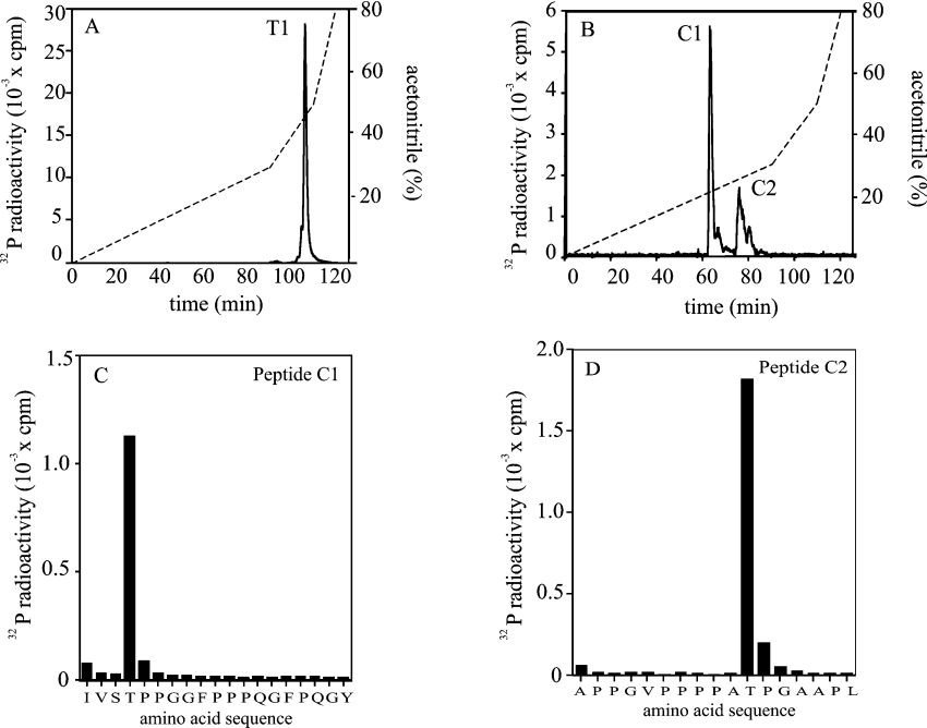
Figure 3. Identification of the residues on DAZAP1 phosphorylated by ERK2.
GST–DAZAP1 was maximally phosphorylated with GST–ERK2, as in Figure 2, and subjected to SDS/PAGE. The band corresponding to 32P-labelled DAZAP1 was revealed by staining with Coomassie Blue, excised and digested with trypsin. (A) The tryptic phosphopeptides were separated by HPLC on a Vydac C18 column equilibrated in 0.1% (v/v) trifluoroacetic acid. The column was developed with an acetonitrile gradient in 0.1% (v/v) trifluoroacetic acid (broken line). Radioactivity is indicated by the continuous line. (B) The major 32P-labelled peptide T1 from (A) was re-digested with chymotrypsin and re-chromatographed on the C18 column as in (A). Peptide C1 (C) and peptide C2 (D) were subjected to solid-phase sequencing to determine the sites of phosphorylation [45]. Amino acid sequences shown are those inferred from MS analysis.

