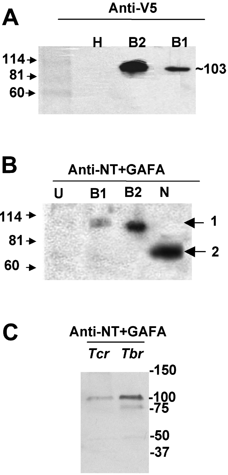
Figure 1. Detection of recombinant T. cruzi PDE proteins using specific antibodies, and Western-blot analysis of TcrPDEBs in T. cruzi trypomastigote lysates.
The Figure shows Western blots of homogenates from: H or U, untransfected HEK-293T cells; B1, HEK-293T cells transfected with TcrPDEB1; B2, HEK-293T cells transfected with TcrPDEB2; N, purified NT+GAF-A fragment with a GST epitope tag expressed in E. coli. The antisera used were: (A) anti-V5; (B) anti-(NT+GAF-A). Molecular-mass markers (shown on the left) are given in kDa; 1, 103 kDa; 2, 72 kDa. (C) Western blot of whole-cell lysates from trypomastigote forms of T. cruzi (Tcr), or bloodstream forms of T. brucei (Tbr) detecting native protein using anti-(NT+GAF-A) antibodies. Molecular-mass markers in kDa are shown on the right.

