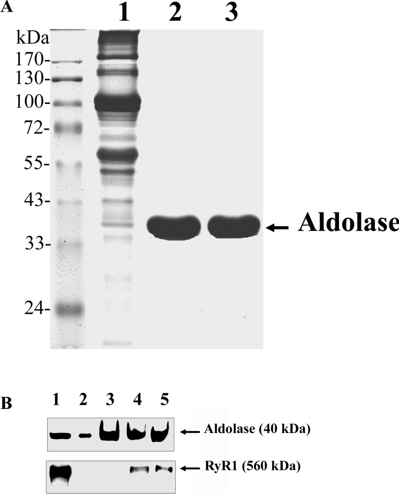
Figure 8. Aldolase-affinity batch-column chromatography.
(A) SR (lane 1), purified aldolase (lane 2) and CNBr-bead-detached aldolase (lane 3) (30 μg of each) were separated by SDS/PAGE (10 % gel) and the gels were stained with Coomassie Blue. (B) Aldolase-affinity beads were incubated with Triton X-100-solubilized native SR. The bound proteins were eluted by SDS sample buffer, separated by SDS/PAGE (7.5 % gel) and subjected to immunoblot analysis with anti-aldolase or anti-RyR antibodies. Lane 1, SR (10 μg); lane 2, purified aldolase (1 μg); lane 3, aldolase-affinity beads alone (2 μg); lane 4, aldolase-affinity bead-bound RyR1 in the absence of DIDS; lane 5, aldolase-affinity bead-bound RyR1 in the presence of 100 μM DIDS.

