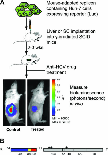
FIG. 1.
(A) Schematic of the HCV replication animal model. Mouse-adapted replicon-containing T7-11 cells expressing a high level of luciferase (Luc) were implanted subcutaneously or directly into the liver parenchyma of γ-irradiated SCID mice. Drug evaluation was performed 2 to 3 weeks after cell implantation. The HCV replication level was monitored quantitatively by measuring the bioluminescence signal using the IVIS imaging system. (B) Structure of the subgenomic replicon rep114/ET derived from pFK I389 LucNS3-3′/5.1 (22) and pFK I341PI Luc NS3-3′/ET (27). 5′, HCV 5 ′ nontranslated region; 3′, 3′ HCV nontranslated region; PVI, poliovirus IRES; Luc, firefly luciferase; Ubi, ubiquitin cleavage site; Neo, neomycin phosphotransferase; EI, encephalomyocarditis virus IRES; *, adaptive mutations E1202G, T1280I, and K1846T.

