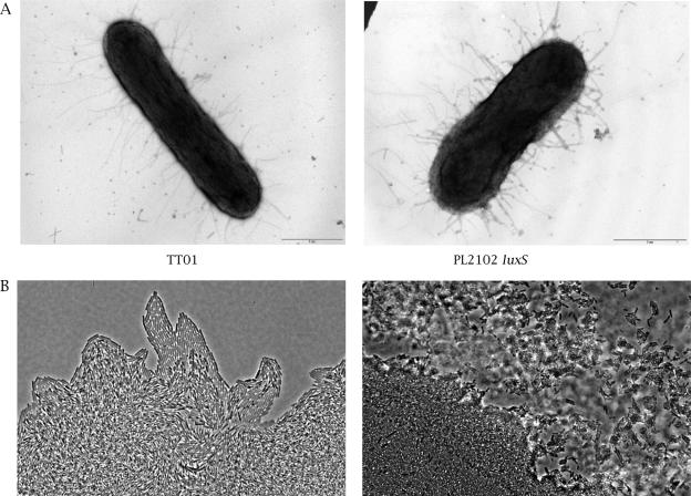
FIG. 5.
Microscopy images. A. Morphology of P. luminescens by electron microscopy. After growth in Schneider medium with 10 μM Na borate, cells were stained with uranyl acetate. B. Light microscopy with phase contrast of expansion zones of twitching motility by slide culture assay, obtained at the interstitial surface between glass cover and medium after 16 h of incubation at 30°C. Magnification, ×40.

