Figure 2.

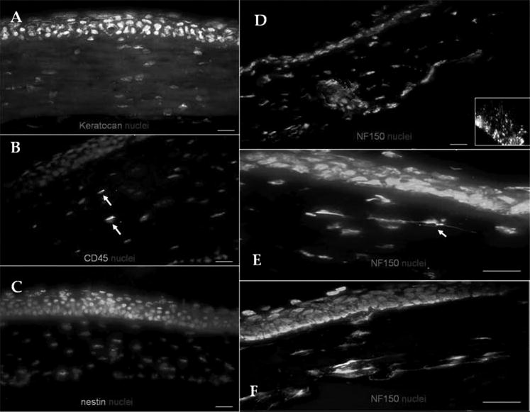
Corneal stroma phenotype in vivo. Noninflamed murine corneas expressed keratocan in the entire stroma (A). Scarce CD45-expressing cells were detected in the stroma (B, arrows). No α-SMA expression was observed (C). NF-150-positive corneal nerve stems entered the posterior sclera and peripheral cornea (D; inset: optic nerve as a positive control). Occasional, nucleated cells expressed NF (arrow) in the 2- (E) and 12- (F) week-old mice. Bar, 25 μm.
