Figure 8.
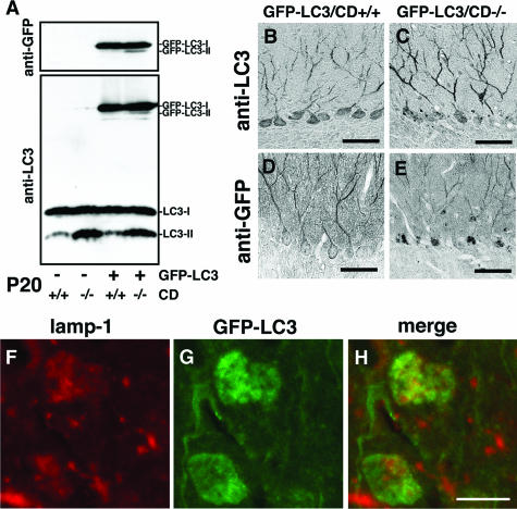
Expression of GFP-LC3 in brains of GFP-LC3/CD−/− mice. A: Molecular forms of GFP-LC3 in brains of GFP-LC3/CD+/+ and GFP-LC3/CD−/− mice at P20. GFP-LC3-I is a major form in both GFP-LC3/CD+/+ and GFP-LC3/CD−/− brains, whereas GFP-LC3-II appears faintly in GFP-LC3/CD+/+ brains and distinctly in GFP-LC3/CD−/− brains when immunostained with either anti-GFP or anti-LC3. Moreover, the quantitative relationship of LC3-II to LC3-I in brains of CD+/+ and CD−/− mice does not differ depending on GFP-LC3 expression. B–E: Immunohistochemical images of GFP-LC3 and LC3 (B and C), or GFP-LC3 (D and E) in cerebellar Purkinje cells of GFP-LC3/CD+/+ (B and D) and GFP-LC3/CD−/− (C and E) mice at P20. Immunoreactivity for LC3-GFP and LC3, that was immunoreacted with anti-LC3, is more distinct and intense in dendrites and bodies of Purkinje cells than that for GFP-LC3 that was immunostained with anti-GFP. Diffuse or fibrillary staining for GFP-LC3 and LC3, or GFP-LC3 is clear in dendrites and bodies of Purkinje cells from a GFP-LC3/CD+/+ mouse, whereas granular staining for them (spheroids, arrows) is observed not only in Purkinje cell bodies but also in their dendrites from a GFP-LC3/CD−/− mouse. F–H: Double-fluorescence images of lamp1 (red color, F) and GFP-LC3 (green, G) in cerebral cortical neurons of GFP-LC3/CD−/− brains at P20. Lamp1 immunoreactivity is often localized in GFP-LC3-positive granules, but a considerable number of GFP-LC3-positive granules are distinct from lamp1 immunoreactivity (H). Scale bars: 50 μm (A–E); 10 μm (F–H).

