Figure 1.
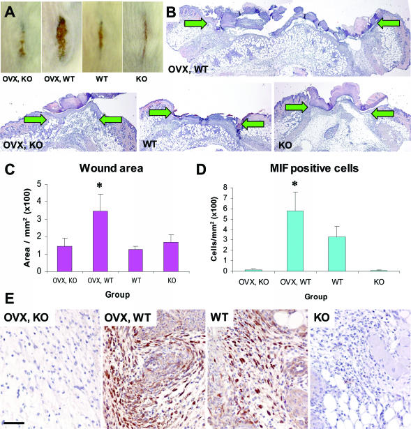
As shown previously MIF is a critical modulator of wound healing, regulated by estrogen in vivo. In mouse, reduced systemic estrogen leads to delayed healing, evident at the gross morphological level (in A, compare OVX, WT with WT) and at the histological level (in B, compare OVX, WT with WT). In the absence of MIF, this effect is abolished (in A, compare OVX, KO with OVX, WT; and in B, compare OVX, KO with OVX, WT). B: Representative hematoxylin and eosin-stained histological sections illustrate the increased wound width (arrows denote the wound edges) and area of wounds (C) from OVX, WT mice compared with other groups; *, t-test P = 0.03. D: Quantification of MIF-positive cells. E: Representative immunohistological MIF localization. D, MIF-null mice lack MIF-positive cells and OVX, WT mouse wounds with reduced systemic estrogen and increased inflammation (A, OVX, WT) have elevated numbers of MIF-positive cells; *, t-test P = 0.0008. E: In intact, WT wounds, MIF localizes predominately to keratinocytes, fibroblasts, and inflammatory cells. In OVX, WT wounds, strong MIF staining is observed in the increased number of inflammatory cells. Bar (E) in A = 5 mm; in B = 370 μm; and in E = 60 μm.

