Figure 5.
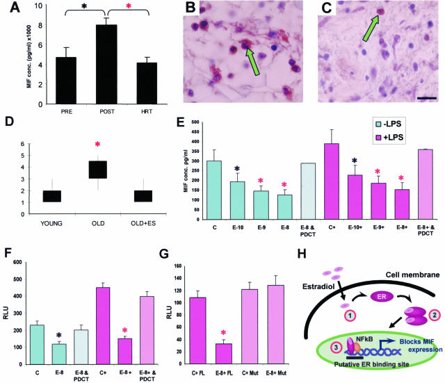
Mechanism of MIF regulation by estrogen in humans. A: Postmenopausal female subjects (45 to 60 years old with low systemic estrogen) have significantly higher systemic MIF levels than premenopausal subjects (35 to 49 years old). MIF levels in female subjects who were postmenopausal and on HRT (47 to 60 years old) were indistinguishable from the premenopausal group. B: Numerous local MIF-positive cells (arrow) in wounds from aged subjects are dramatically reduced by topical estrogen (C; arrow, compare with B). D: MIF immunostaining scored on a scale of 1 to 5 is elevated in elderly subjects and reversed by topical estrogen. E: Human monocyte MIF production, by both resting (−LPS) and LPS-activated (+LPS) cells, is decreased with concurrent estrogen treatment. E-10, E-9, and E-8 = 10−10, 10−9, and 10−8 mol/L estrogen. In addition, treatment of both resting and LPS-activated estrogen-treated monocytes with the NF-κB inhibitor PDCT (60 μmol/L) significantly reversed this estrogen-dependent reduction in MIF levels. F: Human MCF7 ER+ cells transfected with a MIF-promoter luciferase-reporter construct showed increased activity after LPS activation. Estrogen (10−8 mol/L) inhibited this activity, and the effects of estrogen were blocked by PDCT. G: ER-negative JEG3 cells transfected with MIF promoter/reporter and full-length ERα (FL) showed estrogen-dependent (E-8) down-regulation of MIF promoter activation. When a mutant ERα construct (Mut) lacking the AF-1 domain is instead transfected, estrogen is no longer able to inhibit MIF expression. H: Schematic representation of the role of NF-κB in estrogen receptor-dependent activation of MIF. Step 1, estradiol binds to the ER. Step 2, ER dimerizes. Step 3, ER binds to NF-κB. *, t-test P < 0.05; red asterisk, t-test P < 0.01; Error bars = means ± SEM; n = 10–16 (A), 4 (D), 5 (E–G). Bar (C) in B and C = 9 μm.

