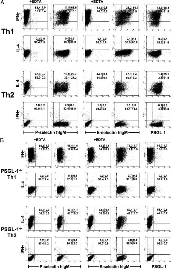
Figure 4.
Analysis of P- and E-selectin ligand expression on wild-type and PSGL-1−/− Th1 and Th2 effector populations. Th1 or Th2 cells were derived from DO11.10 (A) or DO11.PSGL-1−/− (B) mice as in Figure 1 and restimulated with PMA and ionomycin for 6 hours. Monensin was added for the final 4 hours to stop cytokine secretion. T cells were surface-stained with PSGL-1 monoclonal antibody or P- or E-selectin human IgM chimeras. Five mmol/L EDTA was included in the staining media where indicated as a negative control for selectin binding. After surface staining, the cells were fixed, permeabilized, and stained intracellularly for IFN-γ and IL-4. The frequency of IFN-γ+ and IL-4+ cells that were PSGL-1+ and bound P- and E-selectin hIgM was assessed by flow cytometry on the lymphoid gate. Quadrant percentiles of lymphocyte-gated cells are indicated.

