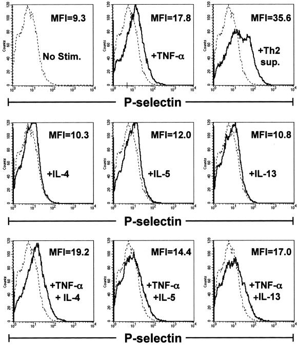
Figure 6.
Th2-derived cytokines do not induce enhanced endothelial cell expression of P-selectin above that induced by TNF-α alone. Confluent bEnd.3 cells were stimulated for 6 hours with nothing (dotted line), Th2 sup, TNF-α (10 ng/ml), IL-4 (20 ng/ml), IL-5 (10 ng/ml), IL-13 (20 ng/ml), or the indicated combinations of cytokines (solid lines). Endothelial cells were recovered, stained with monoclonal antibodies for P-selectin, and analyzed by flow cytometry. In overlay histograms, the mean fluorescence intensity (MFI) for the stimulated condition is given. The MFI of unstimulated bEnd.3 cells = 9.3. Histograms represent 30,000 events.

