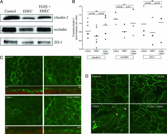
Figure 7.
TGF-β pretreatment prevents EHEC-induced reductions in tight junction protein expression. A: Representative immunoblot showing that TGF-β (10 ng/ml) prevents EHEC (MOI = 100, 16 hours)-induced reduction in claudin-2, occludin, and ZO-1. The events are quantified in B using the integrated pixel intensity of the tight junction protein:claudin-4 bands (n = 3 to 6 separate experiments, each indicated by a different symbol). C: Shown is the epithelial distribution of claudin-2-like immunoreactivity both as en face images and in corresponding composite Z-stack images below each panel (control, Ci; TGF-β, Ti; EHEC, Ei; TGF-β+EHEC, TEi) (images taken in the x-y plane using confocal microscopy; n = 5 monolayers). The diffuse peripheral claudin-2 immunoreactivity in control preparations is lost in EHEC-infected cells and restored to a large extent by TGF-β pretreatment. The z axis images show that although claudin-2 immunoreactivity (green) can occur throughout the cell, the majority occurs in an apical location (nucleus shown as red based on propidium iodide staining) and is located at the tight junction (note intense punctate staining that indicates tight junction regions of adjacent cells). Similarly, and as shown in D, confocal microscopy confirms that TGF-β pretreatment prevents EHEC (MOI = 100, 16 hours)-induced disruption of the distribution of ZO-1. Representative images of T84 cell monolayers immunostained for ZO-1 taken in the x-y plane using confocal scanning laser microscopy (n = 5 to 7 monolayers; arrows denote loss of ZO-1 at the cell periphery and the increased punctate appearance of ZO-1 is highlighted by the asterisk). Original magnifications, ×1260.

