Figure 1.
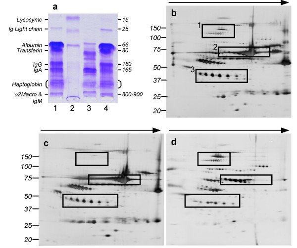
Fractionation of serum protein by IgY12 spin columns followed by 1D and 2D electrophoresis. Panel a: 1D electrophoresis is performed using Hydragel separation kit (Sebia) which allows the detection of major serum proteins including Ig light chains, albumin, transferin, IgG, IgA, Haptoglobin, IgM and alpha-2-macroglobulin. Lane 1: original unfractionated serum; lane 2: molecular weight standard; lane 3: unbound protein fraction (see Data Supplements) and lane 4: IgY12 bound protein fraction. The same amount of total protein was loaded in each lane. Panel b-d: 2D electrophoresis gel of original unfractionated serum (panel b), IgY12 bound proteins (panel c), and Y12 unbound proteins (panel d). Each gel was load with 20 μg of proteins. Some protein spots (box 1 panel b), were present in both the original and the unbound fractions (panel d). Others (box 2, panel a-b) (albumin, haptoglobin.), where retained by the column and recovered in the bound fraction (panel c). Finally, unmasked by the fractionation, some "new" spots were detected only in the unbound fraction (box 3, figure b-d). These results were consistent with the work of Huang et al [18].

