Figure 3.

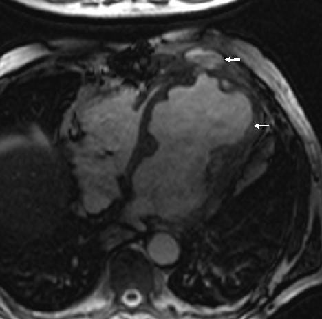
MR scan showing left ventricular aneurysm (arrow) and blood from contained aneurysmal rupture lying under pectoralis minor (small arrow).

MR scan showing left ventricular aneurysm (arrow) and blood from contained aneurysmal rupture lying under pectoralis minor (small arrow).