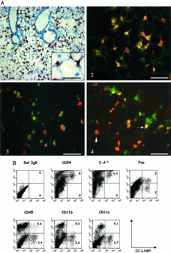
Figure 2.
Characterization of DC-LAMP-expressing lung cells. A: Mouse lung cryosections were stained with anti-DC-LAMP mAb, revealed with AEC substrate, and counterstained with hematoxylin (A1). DC-LAMP is expressed exclusively by a population of alveolar epithelial cells protruding in the lumen (inset). A2 to A4: Double-immunohistofluorescent stainings were performed on mouse lung cryosections. A2: Most lbm180-positive cells (green) co-express DC-LAMP (red). A3: A majority of DC-LAMP-positive cells (red) also co-express CD44v6 (green). A4: Approximately half of DC-LAMP+ cells (red) also express MHC class II (green), but at levels ranging from high (arrow) to low (arrowhead). B: Isolated mouse total lung cells prepared as described in Materials and Methods were surface-stained with various mAbs, permeabilized, and double stained with anti-DC-LAMP mAb. Fluorescence-activated cell sorting data showed that DC-LAMP+ cells express high levels of CD54 and class II (I-Ab) molecules but not CD45, CD11b, or CD11c. Approximately half of the DC-LAMP+ population expresses Fas (CD95). Numbers in the right quadrants indicate percentage of total lung cells. Original magnifications: ×100 (A1); ×400 (A2 to A4). Scale bars, 40 μm.

