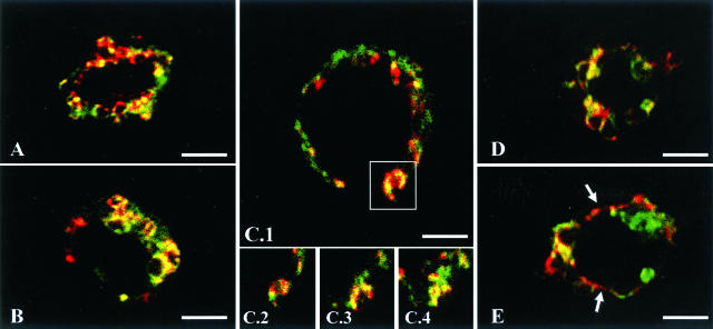
Figure 3.
Subcellular localization of DC-LAMP in PnIIs. Double staining with lbm180 (green) and DC-LAMP (red) was performed on CD45-depleted mouse lung cells immobilized on poly-l-lysine-coated slides. CLM analysis showed co-localization of both proteins in intracellular ring-like organelles (lamellar bodies, LB) (A, B, D). A LB fusing with the plasma membrane is represented in C1 (square), and shown at different levels of the cell (C2 to C4). Cell surface patches of DC-LAMP adjacent to lbm180 were also detected (E, arrows). Original magnifications, ×1000. Scale bars, 3 μm.

