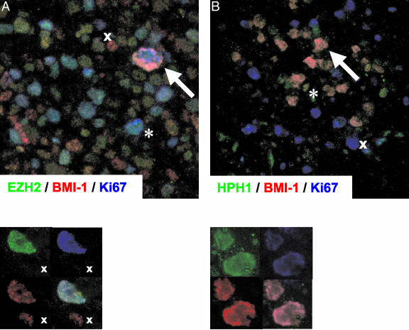
Figure 2.
Immunofluorescent detection of BMI-1 in combination with EZH2 and HPH1 in Ki67POS H/RS cells. A: Triple immunofluorescence staining for BMI-1 (red signal), EZH2 (green signal), and Ki67 (blue signal). Large Ki67POS H/RS cells co-express BMI-1 and EZH2 (arrow) whereas BMI-1- and EZH2 expression are separated in healthy infiltrating cells (x denotes a resting (Ki67NEG) BMI-1POS/EZH2NEG cell; * denotes a dividing (Ki67POS) BMI-1NEG/EZH2POS cell). Lower panel: detail of a single H/RS cell, showing single fluorescence signals (upper left and right, and lower left) and the combination of these signals (lower right). B: Triple immunofluorescence staining for BMI-1(red signal), HPH1 (green signal), and Ki67 (blue signal). Large Ki67POS H/RS cells co-express BMI-1 and HPH1 (arrow) whereas dividing healthy infiltrating cells do not express BMI-1 and HPH1 (x). By contrast, BMI-1 and HPH1 are detectable in resting Ki67NEG healthy infiltrating cells (*). Lower panel: as in A.

